
| • new stuff | |
| • web publishing | |
| • Interactive publications | |
| • photography | |
| • music | |
| • digi video | |
| • Links | |
| • e-Mail mE | |
| • home page | |
|
"It's called a reel change... The movie goes on, and nobody has any idea what just happened. -Tyler Durden.. Fight Club Brain surgery was kind of like having my reel changed... And now my movie rolls on and we'll see how my character arc developes, and where it will end, only God knows... |
|
| My Surgery Report | |
| Take a little look inside the brain of "E".. | |
|
I had a full frontotemporal craniotomy under emergency conditions (which means they cut the whole side of my head open, peeled my scalp back, removed a section of my skull to get in my brain where my bleeding was stopped, the AVM removed, the pools of blood within my skull evacuated, & a resulting hematoma also removed… (I had a massive stroke from all the blood lodging in areas where it had no business being). Click here if you'd like to read the thank you letter I wrote the doc who heroically did the following to me.
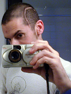
DATE OF Operation: 02/17/2004 The head had been shaved and the horseshoe shaped incision was outlined in the temporoparietal region' above the ear and extending anterior and posterior to this. The head was prepped and draped in the usual manner. The incision was made along the previously mentioned horseshoe shaped curvilinear line. The scalp flap was reflected and the temporalis muscle was incised and the temporalis muscle was reflected from the calvarium. Raney clips were used on the margins and the scalp flap and muscle were wrapped with the moist laparotomy sponge and ~sutured to it and then held backwards. A single bur hole was placed in the temporal area. The dura was separated. The Stryker craniotome was used to cut the bone flap. The bone flap was elevated and the drill holes were made about the margins of the craniotomy and the dura was tacked up to these holes with 4-0 braided nylon. continued, next column. |
continued from bottom of left column: The dura was opened and hinged inferiorly. The temporal lobe and the inferior parietal lobule were thus exposed. The gyri of the temporal lobe were edematous and stretched. In the mid portion of the second temporal gyrus, this was opened and the hematoma was removed. The hematoma extended anteriorly and posteriorly and this was all removed as it was in the sylvian f issure and the substance of the temporal lobe. The hematoma was entirely removed and there was bleeding initially from the cortical surface. This was coagulated. Beneath that, on the brain on the lateral aspect of the clot but on the temporal lobe, there was residual of apparent arteriovenous malformation. This small tangle of blood vessels was coagulated and removed and there was no further bleeding. The area was lavaged and the blood pressure was elevated at 245 mmHg and no bleeding ensued. The area was further lavaged and the temporal lobe was now quite relaxed and pulled away from the calvarium. The dura was then closed with .O nylon. The bone flap was then secured in place with QuickFlap straps. The temporalis muscle was resutured using O-Vicryl and the galea was closed using 3-0 Vicryl and the scalp was closed with staples… (I kept these staples after they were removed and made a maraca out of them… Tee hee… Eric.) Dressings were applied and the patient was returned to the Post anesthesia recovery room. Here's a fun tangent: I'm a bit financially screwed by this, so if you can help out with a donation, well, that will help me continue to donate my talents toward what I think could lead to a better future for all.
Thanks... home page. |

home page | web publishing | Interactive publications | photography | music | digi-video | Links
All content © 2004 ImagiMedia, Inc.. All Rights Reserved.